![]()
Incidence of mycoplasmal pneumonia: Mycoplasmal pneumonia in Japan used to prevail regularly every four years before 1988. Under the National Epidemiological Surveillance of Infectious Diseases (NESID), atypical pneumonia was reported from approximately 2,500 pediatric sentinel clinics before March 1999. In compliance with enforcement of the Law Concerning the Prevention of Infectious Diseases and Medical Care for Patients of Infections (the Infectious Diseases Control Law) in April 1999, the criteria of the disease were changed to mycoplasmal pneumonia including etiological diagnosis, and it is reported as a category V infectious disease from approximately 500 sentinel hospitals (for the criteria of notification, see http://www.mhlw.go.jp/bunya/kenkou/kekkaku-kansenshou11/01-06-24.html).
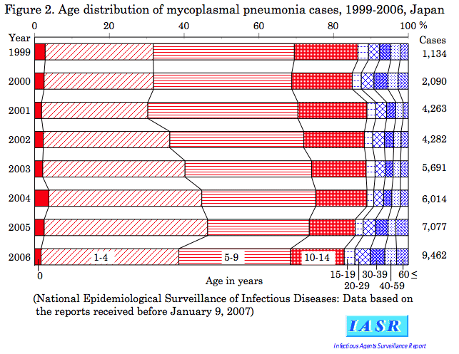
Incidence of atypical pneumonia during 1982-March 1999 and that of mycoplasmal pneumonia after April 1999 are shown in Fig. 1. Large peaks were seen in 1984 and in 1988, but such periodicity has been broken since 1992; from late fall through early spring, small peaks have been recognized regularly since 1991. After 2000, reports of cases per sentinel have tended to increase year after year, and a large increase was seen in 2006, therefore the future trend is worthy of notice. No large epidemic has been seen since 1992, perhaps owing to early diagnosis of mycoplasmal pneumonia and decreased familial infections and outbreaks at schools. Bacterial pneumonia due to Streptococcus pneumoniae or other microorganisms is common among infants and the aged older than 65 years, while mycoplasmal pneumonia is common among pre-school and school children and adolescence age group (Fig. 2). Fig. 3 shows incidence of mycoplasmal pneumonia by prefecture during 2000-2006. Generally, a small number of mycoplasmal pneumonia cases per sentinel occurred in Hokkaido, Shikoku, Kyushu and Okinawa, while in 2006, a large number in Okinawa.
Laboratory diagnosis of mycoplasmal pneumonia: Isolation and culturing of M. pneumoniae from pharyngeal and sputum specimens are conducted only at limited institutions (see p. 38 of this issue). The most common laboratory diagnostic method utilized at present is serodiagnosis. A variety of antibody detection kits are available on the market and simple and rapid tests are applicable. In primary infection of infants and school children within a week after falling ill, negative results may often be obtained. Even if a high antibody titer is obtained with a single serum sample, the possibility of past infection can not be ruled out (see p. 40 of this issue). Recently, detection of M. pneumoniae DNA by PCR has been conducted at gradually increasing institutions (see p. 38 of this issue).
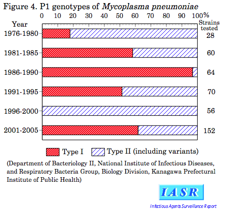
Shift of prevalent M. pneumoniae genotype: M. pneumoniae may be classified into two types according to the different nucleotide sequence of the gene encoding cell-adherent protein (P1). Strains of M. pneumoniae isolated in Japan after 1976 and clinical specimens (pharyngeal swabs and sputum samples) after 2000 were tested by PCR. From the analysis of P1 gene of M. pneumoniae DNA-positive specimens, it has been found that type I and type II organisms have appeared alternatively at certain intervals (see Fig. 4 and p. 38 of this issue). The reason of transposition of the type every 8-10 years can not be explained.
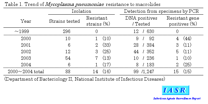
Treatment of mycoplasmal pneumonia: Since mycoplasmal pneumonia resembles clinically chlamydial pneumonia, tetracyclines and macrolides effective on both are generally used before pathogen confirmation, but for children, tetracyclines are not the antibiotics of the first choice because of the possible toxic effects. No strain of M. pneumoniae resistant to macrolides had been found before 1999, while about 15% of M. pneumoniae isolated or detected by PCR were judged as macrolide-resistant after 2000 (Table 1). In cases infected with macrolide-resistant M. pneumoniae , elongation of the febrile period was seen (see p. 41 of this issue). Unless the cases do not undergo worsening, use of macrolides for 7 days (for at least 4 days) is desirable in infants. In addition to macrolides, tetracyclines or fluoroquinolones are used for adults.
Macrolide resistance of M. pneumoniae is due to a point mutation on the 23S rRNA gene of ribosomal 50S subunit. In macrolide-resistant M. pneumoniae strains, mutations of A2063G, A2064G and C2617G were identified on the 23S rRNA domain V (corresponding to the position of A2058, A2059 and A2611, respectively in Escherichia coli ) (see p. 42 of this issue).
Conclusion: In mycoplasmal pneumonia, such new problems as fulminating of adult and aged cases and appearance of resistant organisms have been recognized and treatment based on differential diagnosis of etiological agent is desired. Besides, cycle of epidemics has changed and reports of cases are increasing recently. The cause of such changes is still unknown. Further investigation and pathogen surveillance seem necessary.
 Return to the IASR HomePage
 Return to the IASR HomePage(Japanese)


 Return to the TopPage
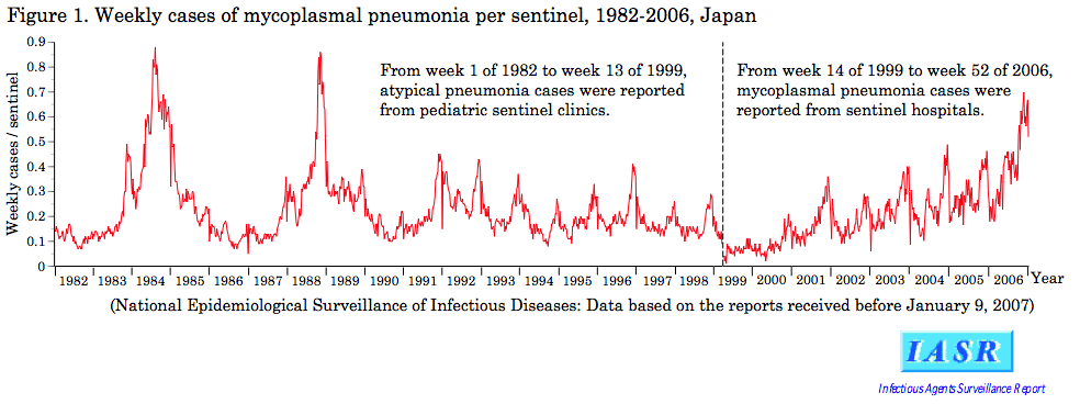