![]()
The Topic of This Month Vol.25 No.4(No.290)
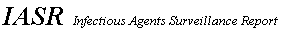
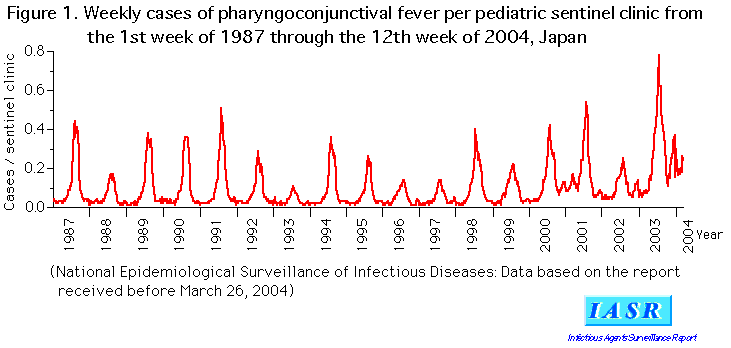
Trend of PCF cases: The PCF cases reported in 2003 from pediatric sentinels of NESID numbered at 40,714 (13.39 per sentinel); they increased at a rate of the highest level of the same week during the past 10 years from the 16th week of 2003, and reached the maximum in the 29th week. They decreased until the 42nd week and turned to increase again after the 43rd week. In 2004, they are still keeping the highest level of the corresponding period in the past (Fig. 1). After 2000, an increasing tendency was seen in winter, which had not been seen before; such tendency has been even more marked later than late autumn of 2003. Yearly incidence per sentinel by prefecture in 2003 (Fig. 2) shows the largest number, 46.0 in Oita prefecture and more than 20.0 in other 11 prefectures. Cases occurring in each age of 1-5 years accounted for 13-16% of all cases and all of them accounted for 75% of all cases in 2003. Such age distribution was similar to that before 2002; in 2004, the proportion of cases of ages under 2 years has increased slightly.
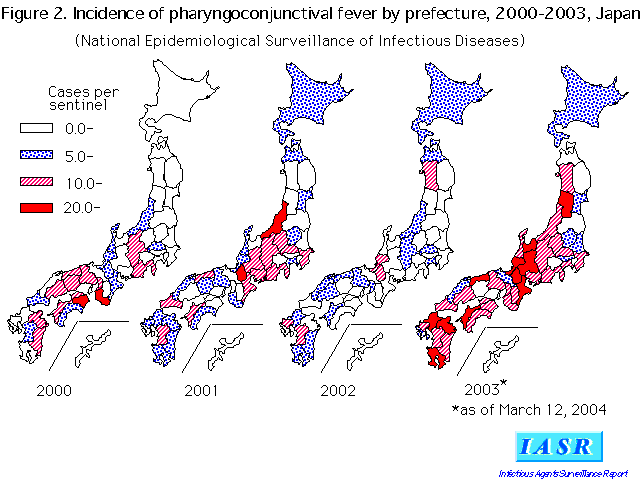
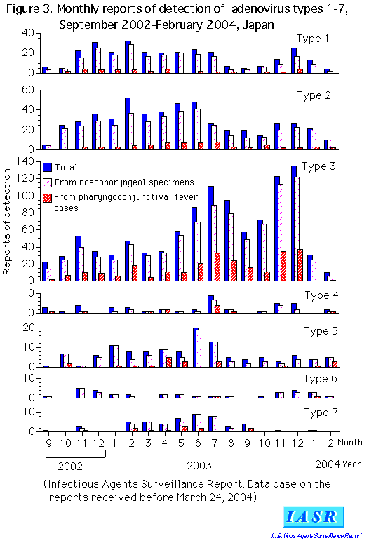
Detection of virus: Specimens, from which adenovirus types 1-7 were detected at prefectural and municipal public health institutes (PHIs) during January-December 2003, are shown in Table 1. Each type was detected mainly from nasopharyngeal swabs (or other nasopharyngeal specimens). A total of 360 cases whose nasopharyngeal swabs yielded virus were diagnosed (see Table 2 on p. 96) as PCF. Adenovirus was detected from 316 of these cases and 209 of them were type 3. The epidemic of adenovirus type 3 in 2003 is characterized by the increase again in winter after the summer peak, as was the case in PCF cases (Fig. 3).
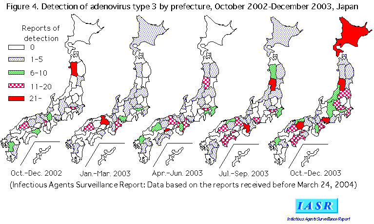
Detection of adenovirus type 3 by prefecture is shown in Fig. 4. During October-December 2002, virus was detected in Akita and other 17 prefectures, during January-Mach 2003 in Hyogo and other 20 prefectures, during April-June in Kagawa and other 27 prefectures, during July- September in Nara, Osaka and Yamagata and other 27 prefectures, and during October-December, when PCF cases increased again, in Yamagata, Okayama, Hokkaido and Aichi and other 29 prefectures.
Type 7, which prevailed during 1995-1998 involving severe cases of pneumonia and fatal cases (see IASR, Vol. 18, No. 4), was reported in 46 cases with a peak in June 2003 (Fig. 3), of which 41 were detected in nasopharyngeal swabs (Table 1). In July 2003, a type 7 outbreak at a dormitory of a sport club of a senior high school in Okayama prefecture was reported (see IASR, Vol. 24, No. 10).
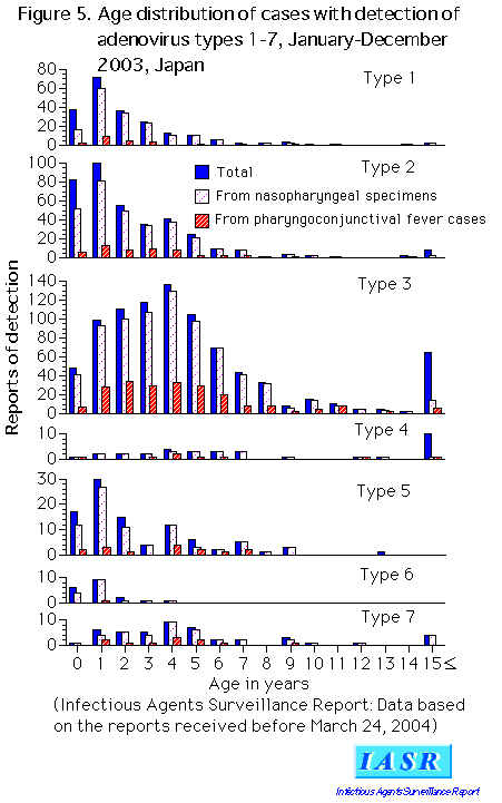
The age distribution of cases, from which adenovirus types 1-7 were detected (Fig. 5), was usual pattern: peaks were shown at 4 years for types 3 and 7 and also at ages older than 15 years, whereas types 1, 2, and 5 were isolated from a low-age group with a peak at one year of age (see IASR. Vol. 21, No. 2).
In recent outbreaks, adenovirus was detected from not only summer PCF cases (see IASR, Vol. 24, Nos. 9 and 10) but also winter outbreaks of common cold or sporadic cases (see p. 96-99 of this issue and IASR, Vol. 24, Nos. 6 and 7). When reports of detection of adenovirus type 3 (Fig. 6) are compared with the occurrence of PCF cases (Fig. 1), detection of adenovirus type 3 from PCF cases (the black area) agreed well to the occurrence of PCF cases. From cases other than PCF (the blank area), a considerable number of adenovirus type 3 was detected in winter seasons.
Conclusions: Adenovirus of not only type 3 but also other types have often been detected from fever of unknown origin, upper respiratory tract inflammation, influenza/influenza-like illness and lower respiratory tract inflammation including pneumonia (Table 2). Therefore, it may be suggested that further attention must be paid to the importance of adenovirus infection as a cause of pediatric and other respiratory diseases.
Adenoviruses of not only type 7 but also of types 3 and 2 may cause severe respiratory diseases, but since the currently available rapid adenovirus antigen-detection kits can not accomplish serotyping, specimens collected from cases of PCF and other respiratory diseases at sentinels for infectious agents surveillance must be subjected to virus isolation and identification at PHIs, the trend of adenovirus serotypes must be captured, and the information obtained must be provided to the medical institutions.
 Return to the IASR HomePage
 Return to the IASR HomePage(Japanese)
